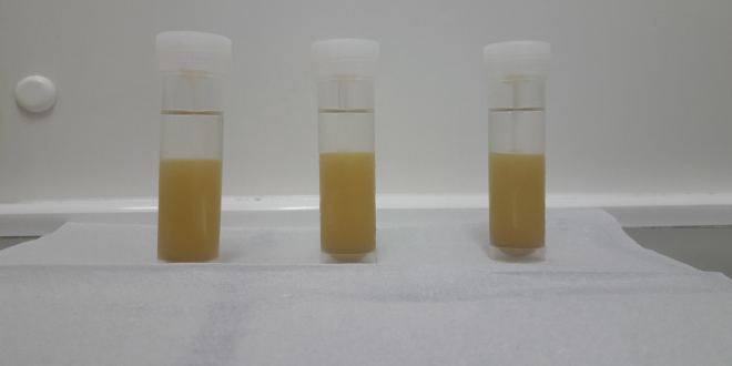
We have investigated the use of aqueous bifasic systems for more effective separation of fecal impurities and intestinal parasites. This study aims at obtaining microscopy slides rich in parasites and free of fecal impurities, which would be an enormous benefit for the automated diagnosis of the parasites.
Lab. of Image Data Science
Projects
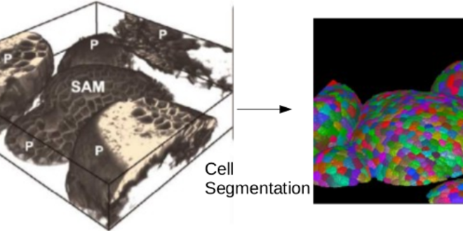
The shoot apical meristem is a network of cells located at the top of plants responsible for all above ground plant growth. Since most of what humans eat, such as grains and fruits, is derived from the shoot, biologists around the world investigate the regulatory mechanisms that drive a healthy growth of the meristem and the crops that depend...
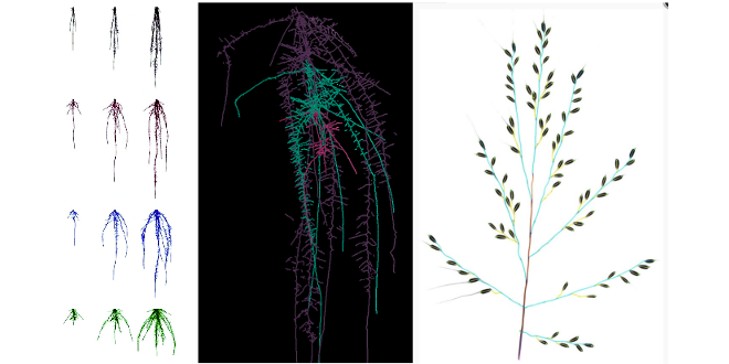
The morfology of plant organs, such as roots, leaves, and seeds, is closely related to relevant properties of the plant. For example, there are evidences that the architecture of a plant root system can indicate its tolerance to acid soils and its capacity to absorb nutrients, such as water and phosphorus. These properties are related to certain genes and...
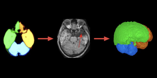
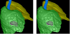
We have developed object shape and texture models to segment human brain structures from MR images. Our approach can separate the left and right hemispheres, and cerebellum without the brainstem, working for normal and abnormal brains. We are currently extending the method to detect brain abnormalities and define abnormal brain asymmetry in association with the study of brain diseases.
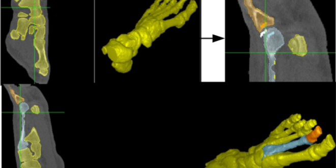
Model-based image segmentation is an important and active field of research in medical imaging. The developed approaches rely on constructing static mathematical representations of an object of interest, by learning the object’s shape from a data set containing previously segmented instances of the object in training images. Those data sets are frequently produced using manual, not interactive, tools, and very...
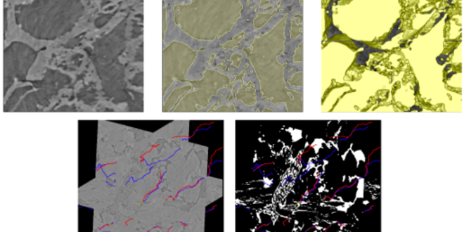
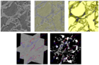
The analysis of sedimentary rock microscopy images allows the characterization of grain morphology in sedimentary petrography. Properties such as grain shape, size, pore tortuosity, and others constitute the basis for such analysis. The estimation of those properties demands the segmentation of the grains (or pores) from the background followed by the mathematical analysis of the segmented objects, a challenging process...
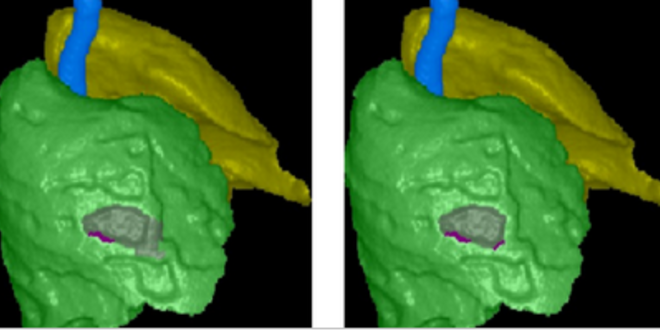
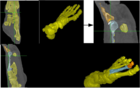
We have developed a fully automated 3D CT-image segmentation method that separates each lung from the trachea-bronchi, in order to study abnormalities on the pleura and interior of the lungs. Lung separation in this case is important to investigate abnormal lung asymmetries. The detection of pleural plaques derived from asbestos is an example. As shown in the cover image, it...
Latest News See all
-
 June 28, 2022 19:16
IC LIDS receives INOVA Inventors Award
-
 February 26, 2020 20:31
Best Student Paper Award at BIOSTEC 2020
-
 March 05, 2020 16:39
DAPI is presented to the International Medical Community