Lab. of Image Data Science
Research
3D Medical Image Segmentation and Analysis
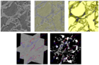
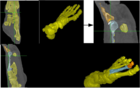
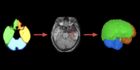
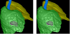
The visualization and quantification of anatomical structures in 3D medical images is paramount to understand the natural course of the disease, plan a treatment, and study the effects of the treatment. This project investigates methods for image segmentation of anatomical structures of the brain and thorax. We are interested in establishing a standard of brain asymmetry from MR images with respect to different measures in order to detect abnormalities in the brain. We are also interested in detecting abnormalities of the pleura and the interior of the lungs.
We have investigated object shape and texture models for 3D medical image segmentation, interactive methods to correct segmentation errors without starting over the process, and methods to learn with the corrections and improve the models.
Projects:
CT Image Analysis of the ThoraxMR Image Analysis of the Brain
Latest News See all
-
 June 28, 2022 19:16
IC LIDS receives INOVA Inventors Award
-
 February 26, 2020 20:31
Best Student Paper Award at BIOSTEC 2020
-
 March 05, 2020 16:39
DAPI is presented to the International Medical Community