Lab. of Image Data Science
Projects
CT Image Analysis of the Thorax
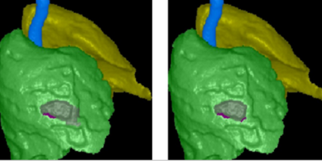
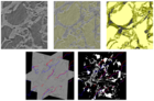
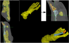
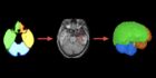
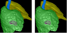
We have developed a fully automated 3D CT-image segmentation method that separates each lung from the trachea-bronchi, in order to study abnormalities on the pleura and interior of the lungs. Lung separation in this case is important to investigate abnormal lung asymmetries. The detection of pleural plaques derived from asbestos is an example. As shown in the cover image, it is possible to visualize them and even segment them interactively for quantification and analysis.
Team:
Alexandre Xavier FalcãoAzael de Melo e Sousa
Elvis Rusnel Capia Quispe
Ericson Bagatin
Klaus Irion
Samuel Botter Martins
Latest News See all
-
 June 28, 2022 19:16
IC LIDS receives INOVA Inventors Award
-
 February 26, 2020 20:31
Best Student Paper Award at BIOSTEC 2020
-
 March 05, 2020 16:39
DAPI is presented to the International Medical Community